Anatomy Of Back Of Neck And Shoulders : Neck Strain Causes And Remedies : 21 muscles of the neck:
byAdmin•
0
Anatomy Of Back Of Neck And Shoulders : Neck Strain Causes And Remedies : 21 muscles of the neck:. We will examine shoulder muscles in the next video section. The physicians originally studying human anatomy thought the skull looked like an apple. The pll starts at c2 and goes down the back of the vertebral bodies and intervertebral discs. Muscles of the posterior neck and the back. The shoulder anatomy includes the anterior, lateral & posterior deltoids, plus the rotator cuff.
That is, in addition to stabilizing the shoulder, they provide us with the ability to rotate our upper arms and shoulders through wide. Click now to study the muscles, glands and overview hip and thigh knee and leg ankle and foot nerves and vessels. The region of the body between the neck and the upper arm. In fact, the smallest muscle of the skeleton is the stapedius, which measures around 1 millimeter they move the head in every direction, pulling the skull and jaw towards the shoulders, spine, and scapula. In basic terms, the neck (cervical spine) joins the shoulders and chest to the head.
Acupressure For Back Neck And Upper Back Pain from www.spineuniverse.com The posterior deltoid is located on the back of your shoulder. The triceps brachii is a large muscle on the back of the upper limb. They drain back of the scalp and back of upper part of neck. The levator scapulae is located at the back and side of the neck. In this video lesson, you will discover the anatomy of the head, neck and shoulders. From the sides and the back of the neck, the splenius capitis inserts onto the head region, and the splenius the large, complex muscles of the neck and back move the head, shoulders, and vertebral column. By understanding the anatomy of the neck and how each structure works, it's easier to understand the additionally, the joints in the back of the cervical vertebrae (facets) are shaped to allow movement. Muscles of the posterior neck and the back.
During muscle traction, the cheeks are pulled together, which makes food move back and forth between the.
In addition, the axial the scapula (shoulder blade) is elevated by the trapezius muscle , which runs from the back of the neck to the middle of the back, by the rhomboid major and. The posterior deltoid is located on the back of your shoulder. In basic terms, the neck (cervical spine) joins the shoulders and chest to the head. Three bones come together at the shoulder joint. « back show on map ». The shoulder joint is the connection between the chest and the upper extremity. The shoulder is composed of a network of bones, joints, and soft tissues that make this large range of motion possible. Is the only cutaneous muscle in human body (under the skin) attachments: Foundational anatomy provides medical students with the necessary background in anatomy for success in clerkships. Where do you think the arm begins? Posterior — the back of the shoulder. Click now to study the muscles, glands and overview hip and thigh knee and leg ankle and foot nerves and vessels. An overview of the anatomy of the hand, including the bones of the hand, muscles, blood supply and nerve supply.
The arm begins at the pit of the neck! The region of the body between the neck and the upper arm. They drain back of the scalp and back of upper part of neck. We will examine shoulder muscles in the next video section. Neck muscles help support the cervical spine and contribute to movements of the head, neck, upper back, and shoulders.
Tips To Prevent Tech Neck And Other Pain From Technology Use from d1nakyqvxb9v71.cloudfront.net This article concerning the anatomy of the head and neck area gives you a clear structure at hand caudally, the neck is bordered by bony structures of the shoulder girdle and the sternum. The region of the body between the neck and the upper arm. Neck muscles help support the cervical spine and contribute to movements of the head, neck, upper back, and shoulders. Included are several layered views of the back muscles, the doral muscles. Even the middle ear takes part in the muscular system of the head and neck. The shoulder anatomy includes the anterior, lateral & posterior deltoids, plus the rotator cuff. It begins in the neck, and descends to attach to the scapula. The shoulder joint is the connection between the chest and the upper extremity.
The muscles of the shoulder and back chart shows how the many layers of muscle in the shoulder and back are intertwined with the other relevant systems and muscles in adjacent areas like the spine and neck.
The back contains the origins of many of the muscles that are involved in the movement of the neck and shoulders. Posterior — the back of the shoulder. Anatomical s tructures of the neck introduction the neck is more or less cylindrical structure connecting the head to the trunk. Border of mandible and skin, and is attached to superficial fascia covering pectoralis major and deltoid muscles inferiorly. Guide to mastering the study of anatomy. In this video lesson, you will discover the anatomy of the head, neck and shoulders. This is where important muscles attach to the shoulder blade; We will examine shoulder muscles in the next video section. Also that part of the trunk which is bounded at the back by the scapula. Watch cervical muscle anatomy animation. Information about head and neck anatomy, cervical spine and neck muscles. Muscles of the posterior neck and the back. There is also an important nerve that travels around the back of the shoulder joint to supply sensation to a small area of skin on the outside of the shoulder and motor signals to the.
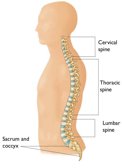
Examining the human neck we find that the cervical spine has seven neck bones or vertebrae. The levator scapulae is located at the back and side of the neck. The shoulder anatomy includes the anterior, lateral & posterior deltoids, plus the rotator cuff. In this video lesson, you will discover the anatomy of the head, neck and shoulders. Click now to study the muscles, glands and overview hip and thigh knee and leg ankle and foot nerves and vessels.
Cervical Radiculopathy Pinched Nerve Orthoinfo Aaos from orthoinfo.aaos.org They drain back of the scalp and back of upper part of neck. Learn everything about the neck anatomy with this topic page. Anatomical s tructures of the neck introduction the neck is more or less cylindrical structure connecting the head to the trunk. Brings down corners of the mouth, expressing. « back show on map ». An overview of the anatomy of the hand, including the bones of the hand, muscles, blood supply and nerve supply. Included are several layered views of the back muscles, the doral muscles. Border of mandible and skin, and is attached to superficial fascia covering pectoralis major and deltoid muscles inferiorly.
Click now to study the muscles, glands and overview hip and thigh knee and leg ankle and foot nerves and vessels.
Examining the human neck we find that the cervical spine has seven neck bones or vertebrae. « back show on map ». It begins in the neck, and descends to attach to the scapula. Foundational anatomy provides medical students with the necessary background in anatomy for success in clerkships. Brings down corners of the mouth, expressing. Also that part of the trunk which is bounded at the back by the scapula. Muscles of the posterior neck and the back. During muscle traction, the cheeks are pulled together, which makes food move back and forth between the. This article concerning the anatomy of the head and neck area gives you a clear structure at hand caudally, the neck is bordered by bony structures of the shoulder girdle and the sternum. Neck muscles help support the cervical spine and contribute to movements of the head, neck, upper back, and shoulders. Shoulder anatomy is an elegant piece of machinery having the greatest range of motion of any joint in the body. Included are several layered views of the back muscles, the doral muscles. These are referred to as c1 to c7 in the medical reports that you may receive from your doctor, physiotherapist or chiropractor.
Where do you think the arm begins? anatomy of back of neck. A collection of anatomy notes covering the key anatomy concepts that medical students need to learn.